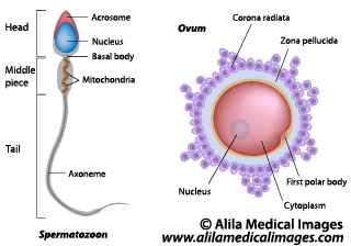
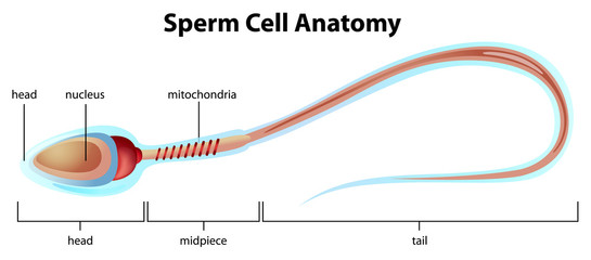
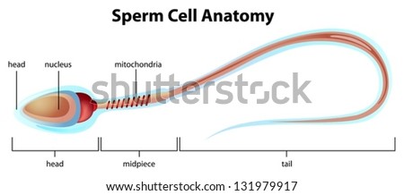
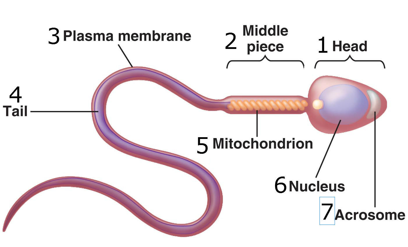
42 sperm cell diagram with labels
Structure of Human Sperm: Check Types of Sperm - Embibe - Embibe Exams Explain the Structure of Human Sperm with Labelled Diagram Fig: Structure of a sperm cell Learn Exam Concepts on Embibe What is the Structure of Sperm? Human sperm is a microscopic structure whose shape is like a tadpole. It has flagella which make it motile. Its diameter is \ (2 - 5 {\rm { \mu m}},\) and its length is \ (60 {\rm { \mu m}}.\) Cells and Reproduction - BBC Bitesize Fertilisation happens when an egg cell meets with a sperm cell and joins with it. The fertilised egg divides to form a ball of cells called an embryo. The embryo attaches to the lining of the uterus.
Spermatogenesis Diagram & Function | What is the Process of Sperm ... Beneath the Sertoli cells are the spermatogonia, which are germ cells that will go through mitosis and ultimately create sperm. In humans, each day, roughly 25 million spermatogonia divide, and...

Sperm cell diagram with labels
Draw a labeled diagram of sperm. - SaralStudy Q:-With a neat diagram explain the 7-celled, 8-nucleate nature of the female gametophyte. Q:-What is oogenesis? Give a brief account of oogenesis. Q:-What is DNA fingerprinting? Mention its application. Q:-Describe the structure of a seminiferous tubule. Q:-What is triple fusion? Where and how does it take place? Draw a labelled diagram of sperm. - Byju's Solution Sperm: It is the male gamete that is released from the male reproductive organ and is responsible for fertilization in human sexual reproduction. The sperm has a few specific parts Head, a Middle piece, and a Tail. The sperm head contains a very little cytoplasm, an elongated haploid nucleus containing chromosomal material. How to draw Sperm Cell || Study of Human Spermatozoon diagram and label ... 'How to draw Sperm Cell || Study of Human Spermatozoon diagram and label the parts' is demonstrated in this video tutorial step by step. Sperm is the male reproductive cell, or gamete, in...
Sperm cell diagram with labels. Testes - Anatomy Pictures and Information - Innerbody Testes. The testes (singular: testis), commonly known as the testicles, are a pair of ovoid glandular organs that are central to the function of the male reproductive system. The testes are responsible for the production of sperm cells and the male sex hormone testosterone. The testes produce as many as 12 trillion sperm in a male's lifetime ... A Labelled Diagram Of Meiosis with Detailed Explanation - BYJUS Diagram for Meiosis. Meiosis is a type of cell division in which a single cell undergoes division twice to produce four haploid daughter cells. The cells produced are known as the sex cells or gametes (sperms and egg). The diagram of meiosis is beneficial for class 10 and 12 and is frequently asked in the examinations. What is a sperm cell like? Its structure, parts and functions - inviTRA Structure and parts of a sperm cell Neck and middle-piece The neck and the middle piece, as the name suggests, are the parts that can be found between the head and the tail. They measure between 6 - 12 microns, a little longer than the head. The width is hardly visible under the microscope. Inside this part are millions of mitochondria. Sperm Cells Images | Free Vectors, Stock Photos & PSD Find & Download Free Graphic Resources for Sperm Cells. 700+ Vectors, Stock Photos & PSD files. Free for commercial use High Quality Images
Human penis - Wikipedia The human penis is an external male intromittent organ that additionally serves as the urinal duct.The main parts are the root (radix); the body (corpus); and the epithelium of the penis including the shaft skin and the foreskin (prepuce) covering the glans penis. Year 7 - Science Revision Guide - Biology CHAUNCY SCHOOL ... Every month an ovum (egg cell) is released from an ovary into the oviduct. This is called OVULATION. If there are sperm cells in the oviduct the ovun may join with one of them. This is called FERTILISATION. The fertilised ovum then travels down to the uterus where it grows Into a baby. The diagram below shows what happens to the ovun after it is Testes: Anatomy and Function, Diagram, Conditions, and Health Tips The epididymis stores sperm cells until they're mature and ready for ejaculation. ... Explore the interactive 3-D diagram below to learn more about the testes. ... (2015). "Off-label" usage ... Sperm Cell Labelled Illustration - Twinkl Sperm Cell Labelled. Use this image now, for FREE! Create your own Sperm Cell Labelled themed poster, display banner, bunting, display lettering, labels, Tolsby frame, story board, colouring sheet, card, bookmark, wordmat and many other classroom essentials in Twinkl Create using this, and thousands of other handcrafted illustrations.
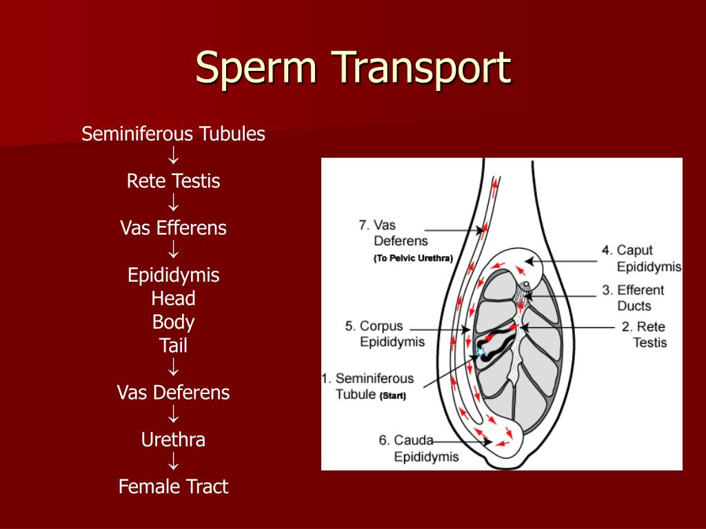
Male reproductive: The Histology Guide - University of Leeds The production of sperm and eggs/ova (gametes) is a procedure called gametogenesis (spermatogenesis and oogenesis). Gametogenesis involves two rounds of meiotic cell division, in which one diploid cell gives rise to 4 haploid cells.. This diagram shows the processes involved in spermatogenesis. The germinal (seminiferous epithelium) of the seminiferous tubules contains spermatogenic cells and ... Sperm Cell Labeled Diagram Stock Vector (Royalty Free) 200461103 ... Frequently used Trendsetter We're seeing significant engagement with this asset. Item ID: 200461103 Sperm Cell Labeled Diagram Formats EPS 6733 × 3563 pixels • 22.4 × 11.9 in • DPI 300 • JPG Show more Contributor j joshya Similar images Assets from the same collection Similar video clips Labeled Sperm Cell Pictures, Images and Stock Photos Browse 15 labeled sperm cell stock photos and images available, or start a new search to explore more stock photos and images. Newest results Receptionist labeling sample in a laboratory Good looking receptionist labeling a urine sample from a male patient in a laboratory front desk Prostate labeled vector illustration. Educational male anatomy... Sperm Diagram Illustrations & Vectors - Dreamstime Download 469 Sperm Diagram Stock Illustrations, Vectors & Clipart for FREE or amazingly low rates! ... Ocean depth zones infographic, vector illustration labeled diagram. Oceanography science educational graphic information. Depth at which sperm whales live and. ... Blue sperm cell vector illustration. 3d fertilisation isolated.
Sperm Diagram Stock Photos, Pictures & Royalty-Free Images - iStock Human Sperm cell Anatomy Penis or Male Reproductive System is a 3D illustration. Comparison between normal and low sperm count Spermatozoons Group Flowing Toward Female Ovum Egg Group of Spermatozoons Flowing Toward Female Ovum Egg Isolated on White Background. Natural Fertilization Process Microscopic View.
Diagram and label sperm cell Diagram | Quizlet Terms in this set (4) Midsection of sperm. contains mitochondria. Sperm nucleus. Contains haploid chromosomes. Acrosome. A vesicle at the tip of a sperm cell that helps the sperm penetrate the egg. Flagellum. A long, whiplike structure that helps a sperm cell to move.
Lymphatic and Immune Systems – Medical Terminology for ... Dec 14, 2021 · Mast cell. Cell found in the skin and the lining of body cells that contains cytoplasmic granules with vasoactive mediators such as histamine. Memory T cells. Long-lived immune cells reserved for future exposure to a pathogen. Monocyte. A type of immune cell that is made in the bone marrow. Mucosa-associated lymphoid tissue (MALT)
Biology: Chapter 10 Assignment Flashcards | Quizlet Review the section "Investigating Life: Heredity and the Hungry Hordes." As indicated in the images below, the dominant allele (R) confers susceptibility to the Bt toxin, and a recessive allele (r) confers resistance to Bt. Suppose that a susceptible male mates with a resistant female.
Draw the diagram of human sperm and label its parts. Write few lines ... Draw the diagram of human sperm and label its parts. Write few lines about it. Medium Solution Verified by Toppr The sperm cells are the haploid gametes which are produced in the male. There are different parts of the sperm cell. (a) Acrosome: This structure contains enzymes used for penetrating the female egg.
Vector — Sperm Cell Labeled Diagram - 123RF Sperm Cell Labeled Diagram Royalty Free Cliparts, Vectors, And Stock Illustration. Image 31015877. Get 10 FREE Images when you get started on our 1 Month-Free Trial. START NOW Vector — Sperm Cell Labeled Diagram Compare Filter Auto-enhance Background removal Size Standard sizes M 2815 x 1490 px L 6733 x 3563 px XL 10099 x 5344 px V SVG or EPS Or
The Mitochondrion - Molecular Biology of the Cell - NCBI ... Thus, the mitochondria in some cells form long moving filaments or chains. In others they remain fixed in one position where they provide ATP directly to a site of unusually high ATP consumption—packed between adjacent myofibrils in a cardiac muscle cell, for example, or wrapped tightly around the flagellum in a sperm (Figure 14-6).
Sperm Under Microscope with Labeled Diagram - AnatomyLearner The principal piece of the sperm's tail The end piece of the sperm's tail Sperm in the epididymis Cyclic events in the seminiferous tubules Sperm under microscope 400x labeled Sperm with 40x and 100x labeled diagram Sperm under the electron microscope Dog sperm under a microscope Abnormalities of the dog sperm Unhealthy sperm under a microscope
labelled diagrams - the sperm cell labelled diagrams funtion of sperm cell table bibliography to the right is a detailed 2D diagram of the sperm cell. there are many parts of a sperm cell. it is extremely small compared to the female egg.
Draw the diagram of the human sperm and label its parts ... - Vedantu It is a microscopic structure, motile in nature, and fertilizes the female gamete, the egg. In mammals, the motile sperm travels with the help of a fluid known as semen. Complete step by step answer: - The human sperm can be divided into the head, the neck, the middle piece, and the tail. - The entire body is enveloped by a plasma membrane.
Specialised animal cells - Cell structure - Edexcel - GCSE Biology ... The tail enables the sperm to swim. Sperm are the smallest cells in the body and millions of them are made. Egg cell. The cytoplasm contains nutrients for the growth of the early embryo. The ...
Sperm Cells - Definition, Function, Structure, Adaptations, Microscopy A normal sperm will have the following characteristics: A smooth oval head - The head of a normally formed sperm has a smooth surface and resembles the shape of an egg The head of the sperm measures 2.5 to 3.5 um in diameter and 4.0 to 5.5 um in length (um=micrometers). This results in a 1.50 to 1.70 length to width ratio
ch 8 mastering biology Flashcards | Quizlet Can you label the phases of the cell cycle? To review a crucial phase of the cell cycle, watch this BioFlix animation: Mitosis. Part A - The cell cycle Drag the pink labels onto the pink targets to identify the two main phases of the cell cycle. Then drag the blue labels onto the blue targets to identify the key stages that occur during those ...

Structure of a sperm cell | Animal Anatomy and Physiology | Pinterest | Homework, Plant cell and ...
cell diagram to label Draw A Diagram Of Human Sperm. Label Only Those Parts Along With Their diagram sarthaks Biology Eleven: Looking At Agiospermae Reproductive Organs. Also biology11lectures.blogspot.com stem monocot plant magnification ovary tissue science microscope section cross leaf lab zea corn mays photobucket botanical biology eleven anatomy
Structure Of Sperm Diagram || Draw Labelled Diagram Of Sperm - YouTube Hello Everyone.Structure Of Sperm Diagram || Draw Labelled Diagram Of Sperm || Class 12 || BiologyStructure Of Sperm Diagram, Draw Labelled Diagram Of Sperm,...
Structure and parts of a sperm cell - invitra.com This labelled diagram shows the structure of a sperm cellin detail, which has the following parts: Head With its spheric shape, it consists of a large nucleus, which at the same time contains an acrosome. The nucleus contains the genetic information and 23 chromosomes.




Post a Comment for "42 sperm cell diagram with labels"