45 simple microscope diagram with labels
Jejunum Histology Slide with Labeled Diagram and Identification Points The villus of the jejunum fold lines with the simple columnar epithelium. Within each villus of the jejunum, you will find the lamina propria. ... In the labeled diagram, I tried to show you all these above-mentioned histological features from the jejunum. Normal jejunum wall histology with diagram. ... In the electron microscope, you will find ... (a) Draw the labelled ray diagram for the formation of image by a ... Click here👆to get an answer to your question ️ (a) Draw the labelled ray diagram for the formation of image by a compound microscope. Derive an expression for its total magnification (or magnifying power), when the final image is formed at the near point.(b) Why both objective and eyepiece of a compound microscope must have short focal lengths?Draw a ray diagram showing the image ...
Microscope Drawing And Label - Painting Valley label microscope diagram compound parts light labeling functions microscopic blank labeled biology microscopy labelled beautiful Compound Microscope ... 496x600 35 0 Parts Of A Compound ... 500x469 27 0 Microscopic Drawing ... 1024x1024 21 4 Download The Diagram... 547x579 17 0 Microscope Labeling ... 270x350 17 0 Microscope Labeling ...

Simple microscope diagram with labels
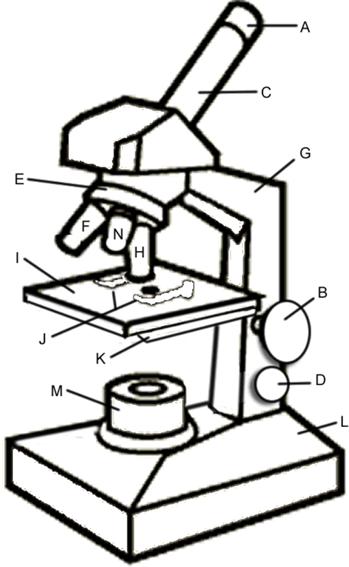
Microscope Parts and Functions With Labeled Diagram and Functions How ... Coarse adjustment: Brings the specimen into general focus. Fine adjustment: Fine tunes the focus and increases the detail of the specimen. Nosepiece: A rotating turret that houses the objective lenses. The viewer spins the nosepiece to select different objective lenses. Objective lenses: One of the most important parts of a compound microscope ... Compound Microscope Parts, Functions, and Labeled Diagram Compound Microscope Parts, Functions, and Labeled Diagram Parts of a Compound Microscope Each part of the compound microscope serves its own unique function, with each being important to the function of the scope as a whole. PDF Parts of a Microscope Printables - Homeschool Creations Label the parts of the microscope. You can use the word bank below to fill in the blanks or cut and paste the words at the bottom. Microscope Created by Jolanthe @ HomeschoolCreations.net. Parts of a eyepiece arm stageclips nosepiece focusing knobs illuminator stage objective lenses
Simple microscope diagram with labels. Parts of a microscope with functions and labeled diagram Structural parts of a microscope and their functions Figure created with biorender.com Figure: Diagram of parts of a microscope There are three structural parts of the microscope i.e. head, base, and arm. Head - This is also known as the body. It carries the optical parts in the upper part of the microscope. Base - It acts as microscopes support. Microscope Types (with labeled diagrams) and Functions Simple microscope labeled diagram Simple microscope functions It is used in industrial applications like: Watchmakers to assemble watches Cloth industry to count the number of threads or fibers in a cloth Jewelers to examine the finer parts of jewelry Miniature artists to examine and build their work Also used to inspect finer details on products Draw a neat (labelled) diagram for the formation of image in a simple ... Draw a neat (labelled) diagram for the formation of image in a simple microscope. Medium Solution Verified by Toppr Light from a light source (mirror) passes through a thin transparent object. A biconvex lens magnifies the size of the object to get an enlarged virtual image. The image is viewed from the other side. Video Explanation Simple Columnar Epithelium: A Labeled Diagram and Functions These form a brush border. They also increase the absorptive surface area of these cells. On a concluding note, simple columnar epithelium has two primary functions of absorption and secretion. In the small intestine, it facilitates the absorption of nutrients. It also secretes mucus, which helps to lubricate, moisten, and protect the surface.
Parts of the Microscope with Labeling (also Free Printouts) Parts of the Microscope with Labeling (also Free Printouts) A microscope is one of the invaluable tools in the laboratory setting. It is used to observe things that cannot be seen by the naked eye. Table of Contents 1. Eyepiece 2. Body tube/Head 3. Turret/Nose piece 4. Objective lenses 5. Knobs (fine and coarse) 6. Stage and stage clips 7. Aperture Microscope labeled diagram - SlideShare Microscope labeled diagram 1. The Microscope Image courtesy of: Microscopehelp.com Basic rules to using the microscope 1. You should always carry a microscope with two hands, one on the arm and the other under the base. 2. You should always start on the lowest power objective lens and should always leave the microscope on the low power lens ... Labeling Microscope Worksheet | Teaching Resources File previews. docx, 300.56 KB. A straightforward worksheet in which students are required to identify the parts of a basic microscope. Tes classic free licence. Microscope, Microscope Parts, Labeled Diagram, and Functions 19 Jan 2022 — There is various type of microscope such as transmission electron microscopes (TEMs), scanning electron microscopes (SEMs), atomic force ...Microscope Parts: Microscope Parts FunctionsObjective lenses: Low-, medium-, and high-po...Light source: Provides light for viewing the spe...Base: Supports the microscope
PDF Biological Diagram Of Simple Microscope With Label Diagram Of Simple Microscope With Label Free Ebooks in PDF format GEOCACHING MERIT BADGE ANSWERS GLENCOE ALGEBRA 1 CHAPTER 7 ANSWERS GETTING''A Level Biology Drawing Skills Booklet OCR June 19th, 2018 - Drawing from a microscope slide8 label and annotate biological specimens students find a Simple Columnar Epithelium Labeled Diagram Simple Columnar Epithelium: A Labeled Diagram and Functions Epithelium is a tissue that lines the internal surface of the body, as well as the internal organs. Simple epithelium is one of the types of epithelium that is divided into simple columnar epithelium, simple squamous epithelium, and simple cuboidal epithelium. Simple Microscope - Parts, Functions, Diagram and Labelling Simple Microscope - Parts, Functions, Diagram and Labelling A microscope is one of the commonly used equipment in a laboratory setting. A microscope is an optical instrument used to magnify an image of a tiny object; objects that are not visible to the human eyes. Table of Contents The common types of microscopes are: What is a Simple microscope? Simple Microscope - Definition, Types, Working Principle & Formula A simple microscope consists of a convex lens of a short focal length. The below figure shows the ray diagram which subsequently forms the image of an object (or we can say a source of light). (Image will be Updated soon) F is the focal length of the lens. An object is placed between the focal length and the centre of the curvature.

Chapter 1, Page 12 - HistologyOLM
Simple microscope | Fun Science Principle of Simple Microscope. A simple microscope works on the principle that when a tiny object is placed within its focus, a virtual, erect and magnified image of the object is formed at the least distance of distinct vision from the eye held close to the lens. Working of Simple Microscope. The ray diagram to show the working of simple ...
Microscope Coloring - ClipArt Best - ClipArt Best
A Study of the Microscope and its Functions With a Labeled Diagram To better understand the structure and function of a microscope, we need to take a look at the labeled microscope diagrams of the compound and electron microscope. These diagrams clearly explain the functioning of the microscopes along with their respective parts. Man's curiosity has led to great inventions. The microscope is one of them.

anatomyforme: 2008-04-06
Compound Microscope Parts - Labeled Diagram and their Functions - Rs ... There are three major structural parts of a compound microscope. The head includes the upper part of the microscope, which houses the most critical optical components, and the eyepiece tube of the microscope. The base acts as the foundation of microscopes and houses the illuminator. The arm connects between the base and the head parts.
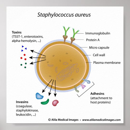
Staphylococcus aureus bacterium labeled diagram. poster | Zazzle.com
Simple Microscope Definition, Magnification, Parts And Uses To make a simple microscope with the help of water. Apparatus Required A glass of water Fuse wire Object to view (newspaper works well due to its fine print) Procedure Make a loop of the fuse wire around 2 mm wide. Dip it in water so that a drop is made in the loop. Hold it near to your eye and take a close look at the object you have chosen.
Flatworms
Parts of a Simple Microscope - Labeled (with diagrams) Parts of a Simple Microscope - Labeled (with diagrams) A simple microscope is a very first type of microscope ever created. It consists of simple parts and performs simple functions. Although there are now many advanced microscope types, some applications may still demand the use of a simple microscope.
Print Anatomy & Physiology: The Integumentary System flashcards | Easy Notecards
Label the microscope — Science Learning Hub All microscopes share features in common. In this interactive, you can label the different parts of a microscope. Use this with the Microscope parts activity to help students identify and label the main parts of a microscope and then describe their functions. Drag and drop the text labels onto the microscope diagram.
Post a Comment for "45 simple microscope diagram with labels"